JPID - Vol 04 - Issue 02

3D BIO PRINTING IN MAXILLOFACIAL REGION

*Sarath S S, **Ajithkumar K; *Abhilash B, *Jyothi K N, ***Vidya K G; ****Prasanth V
* Assistant professor, **Professor and HOD, Department of OMFS, Government Dental college, Thiruvananthapuram, ***Assistant professor, Department of Conservative Dentistry and Endodontics, Government Dental College, Kozhikode, ***Associate professor, Department of Prosthodontics Government Dental college, Alappuzha | Corresponding Author: Dr Sarath S, E-mail: sarathgolfer@gmail.com

Abstract:

Loss of tissue in maxilliofacial region is so common due to various reasons like trauma, cancer surgeries etc…With the arrival of tissue engineering methods viable tissues can be “cultivated” in the lab and can be used for curing the defects, novel method of 3D bioprinting is the new hope in tissue engineering.

Introduction

The ability to print biological tissues opposite to traditional 3D plastic and metal printing has resulted in the birth of the new bio printing and Tissue Engineering research field. 3D bio printing is a computer-aided deposition of cells, biomaterials and biomolecules. The advantage of 3D bio printing compared to traditional tissue engineering is assembling cells, biomaterials and biomolecules in a spatially controlled manner to reproduce native tissue. In the future, due to highresolution characteristic of printing technology with novel printable biocompatible materials or ‘inks’, autologous tissue will be 3D printed with macroand micro-architecture for reconstruction. The focus is by controlling the micro and macrostructures to replicate complex native-like tissue architecture more reliably than by conventional methods. The wide synergy of research on biomaterials and on 3D bio printing may enable restoring the form and functional reconstruction of OMF anatomy in the near future. 3D bio printing avoids donor site complications and immunosuppression. The main obstacles for wider use for 3D bio printing are related to biology, technology and regulatory issues.

Traditional 3D printing is relatively simple and can be performed by the home computer using the proper software. For medical use, 3D digital data are acquired from computed tomography, magnetic resonance imaging, or laser scanning. These data can be manipulated by CAD–CAM software and be converted into Stereolithographic format for printing. Fabrication of solid bio model is carried out under the computer guidance to accurately and in a controlled manner deposit biological materials in a layer-by-layer fashion. 3D bio printer uses a nozzle to deposit biomaterials and cells according to xyz-axis to create the structure required. Fabricated solid model is then cultured in a bioreactor under specific conditions to produce specific and designed tissue engineered vital tissue.

BioInk, How is it produced?

Cells derived from patients preferably stem cells are cultured in optimum conditions. These cells are provided with adequate growth factors enabling them to grow and multiply. When they have multiplied to enough numbers they are collected formed into spheroids and loaded into a cartridge to create a bio ink.


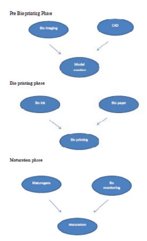
Bio printing involves three phases

  1. Pre Bio printing Phase: In this phase choosing of material for bioprinting and model creation is done. Biopsy of the required organ is done and cells are mixed with special liquefied medium.
  2. Bio printing Phase: In this phase bio inks are loaded into specific catridges and patient’s scan is analysed for the extent of the defect. According to the defect bioinks are deposited to get the appropriate “printed tissue”.
  3. Post processing phase: In this phase printed construct is transferred into a bioreactor, which could be a simple incubator or a specifically designed culture environment that enables the control of environmental variables that affect biological processes. The nutrients and maturogens are made available for the tissue.

3D Bioprinting techniques

  1. Stereolithography is regarded as the first 3D printing technique. It uses a laser beam to polymerize photocurable resin layer by layer. It was initially developed to create high-resolution rapid prototypes and therefore, due to lack of biocompatible resins, has limited utility in biofabrication. However, improvements in biocompatibility and biodegradation of resins make stereolithography a promising bioprinting technology of the future. It can replicate high resolution and enables to create complex shapes and microstructure

    Eg: Photosolidification

  2. Extrusion-based bioprinting is based on the dispense of viscous bioink with biomaterials, biomolecules and cells through a nozzle. Viscous liquid or molten material extruded through nozzle as a continuous strand of individual dot. After printing, the loose model can be solidified layer by layer. Cell viability in the printed ‘tissue’ seems to be as high as 90% in spite of forces and higher temperatures. Material viscosity and potential forleaks can affect resolution. It also provide limited mechanical stiffness

    Eg: Fused deposition modelling

  3. Laser-assisted bioprinting uses laser beam guided direct writing to induce the transfer of material from a source film onto a nearby receptor substrate in the form of a microdroplet. Apart from the doubts of minor cell viability compared to other 3D techniques, laser-assisted bioprinting has been shown to print mammalian cells without affecting their function. This technique can also provide High resolution and it is compatible with wide range of biomaterial viscosities. Major disadvantage is low cell viability.

    Eg: Laser-guided direct writing

  4. Inkjet printing uses microdroplets of cells for printing of 3D high-resolution models. Some of the major drawbacks include cell viability at higher temperatures and pressures during the printing process that may lead to low cell density within the 3D biomodel. The advantages of this technique include the ability to combine multiple cell types and high resolution to print complex structures. Present research on this bioprinting suggests it as a promising technology even though chances of cell death is there.

    Eg: Thermal 3D inkjet bio printing

  5. The nanobioprinting uses nanoscale surface scaffolds to either increase cell-to-matrix interactions or incorporate nanoparticles into bioinks to noninvasively manipulate and track cells within tissue-engineered structures, for example, adding magnetic iron oxide to ‘bioink’ and using magnet as an external manipulation. This technique allows scaffold surface modification on the nanoscale for additional functionality still at present little is known about the cell behaviour

    Eg: Dip pen nanolithography

    At present, the main challenges of 3D bioprinting technologies are related to (1) biological, (2) technological and (3) regulatory aspects.

    From biological perspectives, not only depositing cells, scaffolds and biomolecules in a spatially controlled manner is sufficient to create durable native-like tissue. A critical step is the transition of mechanically weak 3D bio printed neo-tissue constructs to native-like functional tissue that is transplantable into human. This development leading to functional tissue takes place invitro bioreactor-based culture by using a various physiological conditions and growth factors and their combinations. It may also take place in vivo through the implantation of the 3D bioprinted construct. The challenge is also a lack of vasculature and nutrition due to the size of Tissue Engineered constructs. Printing complex composite tissue has additional challenges, such as long bio-manufacture times which may result in reduction of cell viability, and reduction in cellular dedifferentiation with loss of regenerative potential.

    From technological perspectives, two obstacles are still unsolved. The microstructure of bioprinted constructs and the optimal printable material remain the major research focus for printing complex biological structures. Detailed and accurate microstructure not only increases similarity to native architecture but can also enable physiological pore size and interconnectivity, which is in turn important when considering that diffusion distances of over 400–500 μm limit oxygen and nutrient transport to cells. Currently, 3D printing techniques are diverse in properties; some, like stereolithography, provide high resolutions but are limited in appropriate biomaterials and low cell viabilities.

    Clinical use of bioprinted structures includes ensuring the safety particularly with regard to growth potentials and practicalities like stem cell banks, upscaling, sterility and storage of tissue-engineered constructs. 3D bioprinted materials need to comply with good manufacturing practice regulations and need to be approved by regulatory authorities. One of the main difficulties will be to standardize, validate and monitor 3D bioprinting process from planning and design to manufacturing phase. Bioprinting an utmost intrinsically variable patient-specific process and hence, extremely troublesome. Several bioprinting technologies are promising, but because each tissue currently requires a particular technology, the printing of multicellular tissue constructs is difficult and the mechanical stability of current ‘bioinks’ is not satisfactory for reconstruction.

CONCLUSION

Although anatomical and functional bone identical to original jawbone cannot be produced at the present, there is good evidence that Tissue Engineered bone identical to missing bone part will be available in the near future. This is an area that could revolutionize the oral and maxillofacial reconstruction in near future.

REFERENCES

  1. Tissue Engineering in Oral and Maxillofacial Surgery, Riitta Seppänen-Kaijansinkko
  2. Mironov V. The Second International Workshop on bioprinting, biopatterning and bioassembly. Expert OpinBiolTher. 2005;5(8):1111–5.
  3. Vacanti CA. The history of tissue engineering. J Cell Mol Med. 2006;10:569–76.
  4. Ingber DE. Mechanical control of tissue growth: function follows form. PNAS. 2005;102(33):11571–2.
  5. Dowler C. Automatic model building cuts design time, costs. Plast Eng. 1989;45:43–5.
  6. Fisher JP, Dean D, Mikos AG, et al. Photo crosslinking characteristics and mechanical properties of diethyl fumarate/poly (propylene fumarate) biomaterials. Biomaterials. 2002;23:4333–43.
  7. Melchels FP, Feijen J, Grijpma DW, et al. A poly (D, L-lactide) resin for the preparation of tissue engineering scaffolds by stereolithography. Biomaterials. 2009;30:3801–9.
  8. Chan V, Zorlutuna P, Jeong JH, et al. Three dimensional photopatterning of hydrogels using stereolithography for long-term cell encapsulation. Lab Chip. 2010;10:206–70.
  9. Jakab K, Norotte C, Damon B, et al. Tissue engineering by selfassembly of cells printed into topologically defined structures. Tissue Eng A. 2008;14:413–21.

JPID – The journal of Prosthetic and Implant Dentistry / Volume 4 Issue 2 / Jan–Apr 2021

Copyright © 2017-2023 Indian Prosthodontic Society Kerala State Branch | IPSKERALA.COM | Powered by Dentaura.Com