Neuroplasticity has become increasingly relevant in
understanding how the brain adapts to prosthodontic
rehabilitation. This narrative review examines
current evidence on how dentures, implant
supported prostheses, and occlusal splints influence
cortical reorganization and the restoration of
sensory–motor function. A literature search of studies
published between 2008 and 2025 was conducted
through PubMed and MEDLINE, with supporting
evidence obtained frompeer-reviewed publications
outside these databases. Findings show that oral
rehabilitation reactivates somatosensory and motor
cortices, improving mastication, proprioception,
and functional control, with particularly significant
benefits in elderly patients. Conversely, maladaptive
plasticity contributes to temporomandibular
disorders, which can be modified through occlusal
stabilization and neuromuscular therapies.
Emerging innovationssuch as digital prostheses,
neuromuscular stimulation, and sensor-based
systems, further support adaptive cortical change.
Understanding these neural mechanisms broadens
the prosthodontic perspective, highlighting that
successful rehabilitation restores both oral structures
and the brain’s capacity to relearn function.
Key words: neuroplasticity, corticalreorganization, osseoperception, neural adaptation, oral rehabilitation
Prosthodontics focuses on restoring oral
function, appearance, and patient comfort
through the replacement of missing teeth and
surrounding structures. Its purpose extends
beyond mechanical reconstruction and it
seeks to reestablish neuromuscular harmony
and sensory perception within the masticatory
system. Tooth loss disrupts the complex network
of proprioceptive feedback from the periodontal
ligament, oral mucosa, and masticatory
muscles, leading to altered bite control, reduced
masticatory efficiency, and even changes in cortical processing related to oral function.
Neuroplasticity, defined as the brain’s ability to
reorganize its structure and function in response
to sensory and functional stimuli, plays a
key role in adapting to these changes. Once
believed to occur only in childhood, studies
have now established that the adult brain
retains a remarkable capacity for structural and
functional remodeling, enabling recovery of
lost or altered functions1. Early thinkers such as
William James and Jerzy Konorski introduced the
concept of neural adaptability, which has since
been validated through modern imaging and
neurophysiological research. In prosthodontics,
this adaptability manifests as cortical
reorganization following denture or implant
rehabilitation, where neuroimaging studies
demonstrate reactivation of the primary motor
and somatosensory cortices after prosthesis
use2. These neural adjustments contribute to
improved coordination, proprioception, and
masticatory control. The concept of neuroplastic
prosthodontics
has therefore emerged,
emphasizing that prosthetic treatment is not
merely a structural replacement but a process of
neurosensory training. Repeated sensory-motor
activities such as chewing, occlusal contact,
and proprioceptive feedback reinforce neural
pathways, enhancing adaptation and long-term
oral function.1,2
Neuroplasticity is the brain’s intrinsic ability to
reorganize its structure and function in response
to sensory alterations, motor demands, or injury.
It enables the nervous system to form new
synaptic connections and reorganize existing
neural pathways to maintain or recover lost
function. In prosthodontics, this adaptability
is of particular significance, as tooth loss and
subsequent prosthetic rehabilitation alter oral
sensory feedback and motor coordination, requiring the brain to relearn and refine patterns
of mastication and proprioception. Two principal
types
of
neuroplasticity
are
recognized:
structural plasticity, involving physical changes
in dendritic connections and synapse formation,
and functional plasticity, which reflects variations
in neural activity and synaptic efficiency within
established circuits. These complementary
processes enable the cortex to adapt continuously
to changing peripheral inputs.2,3
Cortical plasticity, a subset of neuroplasticity,
occurs when the cortical representation of a
body part changes following sensory or motor
modification. Loss of natural teeth reduces
activity in cortical regions responsible for oral
sensation and motor control. Studies using
functional magnetic resonance imaging (fMRI)
have shown that rehabilitation with dentures
or implant-supported prostheses restores and
enhances activity within the primary motor and
somatosensory cortices through mechanisms
of cortical remapping and increased regional
cerebral blood flow. Such cortical reorganization
supports improvements in masticatory efficiency,
bite control, and oral perception, highlighting
the neural basis of functional recovery in
prosthodontic treatment.4
Tooth loss and reduced mastication profoundly
affect both oral and cognitive functions,
particularly in elderly individuals. Edentulism
leads to decreased sensory input from periodontal
and oral mechanoreceptors, resulting in reduced
stimulation of the somatosensory and motor
cortices5. This deprivation can cause cortical
reorganization and diminished activity in brain
regions responsible for memory, attention, and
coordination. The loss of afferent signals from
the masticatory system, termed deafferentation,
alters neural integration between the oral cavity
and the central nervous system, which may contribute to cognitive decline and reduced
motor precision6. In older adults, edentulism
has been strongly associated with a higher risk
of dementia and Alzheimer’s disease. Studies
have shown that reduced chewing activity lowers
cerebral blood flow and oxygenation in the
prefrontal cortex and hippocampusregions which
are vital for memory and executive function. This
relationship supports the emerging concept of a
brain–stomatognathic axis, which describes the
close neural connection between mastication
and brain performance. Chronic loss of oral
input accelerates hippocampal degeneration
and impairs cognitive processing in contrast
to restoring masticatory function which may
reverse or mitigate these effects.7
Oral rehabilitation through dentures or implant
supported
prostheses
has
demonstrated
measurable improvement in brain function.
Functional MRI studies show that mastication
with dentures reactivates the prefrontal cortex
and hippocampus, enhancing cognitive tasks
related to attention and recall. Implant-retained
overdentures, in particular, produce stronger
cortical responses and higher Mini-Mental State
Examination (MMSE) scores than conventional
complete dentures, suggesting superior sensory
feedback and cortical stimulation6. Another
clinical study also reported that rehabilitation
of masticatory function in older adults led to
significant improvement in episodic memory and
executive performance over one year of follow
up7. These findings emphasises the concept that
oral rehabilitation is not solely a mechanical
restoration but a neurosensory reactivation
process, by reinstating sensory feedback,
occlusal stability, and masticatory rhythm where
theprosthodontic treatment can stimulate cortical
reorganization and supports cognitive health.
Restoring masticatory efficiency can therefore
be considered a modifiable factor in preserving
brain function, particularly in populations at risk of age-related cognitive decline.5,8
The process of adaptation following prosthodontic treatment demonstrates the brain’s ability to reorganize in response to altered sensory and motor inputs from the oral cavity. Tooth loss leads to reduced afferent stimulation from periodontal and oral mechanoreceptors, while prosthetic replacement restores this input through alternate feedback mechanisms. These neural adjustments are central to functional rehabilitation and are supported by both electrophysiological and neuroimaging evidence.1
Temporomandibular disorders (TMD) reflect
maladaptive neuroplasticity, where persistent
nociceptive input from the joint and masticatory
muscles alters cortical organization and motor
control. Repeated pain input strengthens
maladaptive neural pathways, causing the
brain to “learn” dysfunctional patterns that
maintain pain, muscle overactivity, and altered
jaw movements even after the original cause has
resolved. Functional MRI and EEG studies show
hyperactivation of the primary somatosensory
and motor cortices and increased limbic
activity, consistent with central sensitization14. Prolonged TMD disrupts precise cortical maps
of jaw musculature, impairing coordination
and bite regulation5. Disturbed occlusal and
proprioceptive feedback further modifies
cerebral blood flow and cortical activity, similar to
neural adaptation seen after oral rehabilitation.6
Occlusal splints act as a neurophysiologic
stabilizer by re-establishing balanced sensory
input and reducing abnormal muscle activity.
Splint therapy modulates trigeminal afferents,
restores cortical symmetry, and promotes
normalization of sensorimotor activation.14 When
combined with physiotherapy or biofeedback,
these interventions utilize adaptive plasticity to
reduce pain and re-establish coordinated motor
control.
Neuromuscular Training and Digital Rehabilitation
Neuromuscular stimulation and targeted
masticatory exercises have been shown to
enhance cortical adaptation after prosthodontic
treatment. EMG-guided training, biofeedback,
and repetitive chewing routines improve
trigeminal–motor coordination and shorten the
learning phase for new dentures or implants.
Sylvana A M (2024) reported that neuromuscular
stimulation devices strengthen facial and
masticatory muscle activity, supporting cortical
reorganization and faster prosthetic adaptation.
Parallel progress in digital denture design,
especially CAD/CAM fabrication, provides
precise occlusal balance and fit, minimizing
irregular sensory input and promoting stable
cortical mapping during rehabilitation.15
Smart Prostheses and Neuro-Integrated
Technologies
Advances in sensor-embedded and AI-assisted
prostheses are redefining oral rehabilitation.
Pandey et al. (2025) described implant systems with intraoral pressure sensors capable
of delivering tactile feedback that mimics
periodontal sensation, facilitating cortical
remapping and improved proprioception. These
smartprostheses use adaptive AI algorithms to
analyzeocclusal force and mastication rhythm
in real time, guiding patient-specific training
and optimizing neural adaptation. Future
neuro-integrated interfaces and braincomputer
communication models hold potential for
bidirectional exchange between prostheses and
the central nervous system, enabling voluntary
modulation of bite force and occlusal precision.16
Neuroplasticity provides the biological basis
for how prosthodontic treatment restores
function after tooth loss. Edentulism disrupts
afferent input from periodontal and oral
mechanoreceptors,
producing cortical
reorganization and reduced sensorimotor
precision. Oral rehabilitation, whether through
complete dentures or implant-supported
prosthesisreactivates
the somatosensory
and motor cortices, improving masticatory
coordination, bite force, and proprioceptive
awareness9,10. Functional imaging confirms that
cortical activation increases as patients adapt to
new prostheses, showing that rehabilitation is a
neurobiological as well as mechanical process1.
In older adults, oral rehabilitation contributes
not only to chewing efficiency but also to
cognitive maintenance. Reduced mastication
in the elderly has been linked to diminished
hippocampal activity and accelerated cognitive
decline. Re-establishing masticatory function
through well-adapted prostheses restores
cerebral perfusion and promotes activation of
prefrontal and hippocampal areas6,17. These
effects demonstrate that prosthodontic care in
the elderly promotes both oral and neural health,
limiting age-associated cortical regression.
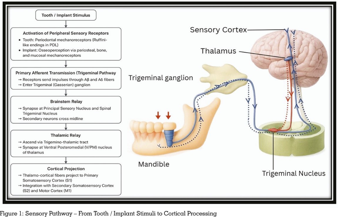
Implant rehabilitation offers further neuroplastic
benefit through osseoperception
where
mechanical signals are transmitted via bone
and periosteal mechanoreceptors that evoke
tactile feedback closely resembling to natural
dentition. Functional MRI studies demonstrate
progressive normalization of cortical activity
following implant loading, suggesting sensory
substitution and new pathway formation.
Mechanotransduction at the implant interface
therefore contributes directly to cortical re
adaptation.11,12
Conversely, temporomandibular disorders
(TMD) illustrate maladaptive neuroplasticity.
Persistent nociceptive input induces cortical
hyperexcitability and disorganized motor
control, reinforcing pain cycles14. Occlusal splints
and physiotherapy correct these maladaptive
circuits by stabilizing occlusal input and
restoring symmetrical sensorimotor activation.
Similar adaptive responses have been observed
in patients with neuromuscular disorders such
as Parkinson’s disease, where rehabilitative
prostheses improve oral motor coordination and
cortical responsiveness.5,17
Prosthodontics is entering an era where
rehabilitation is viewed as guided cortical
retraining
rather than mere structural
replacement. Approaches such as neuromuscular
stimulation, EMG-guided exercises, and
digitally fabricated prostheses promote adaptive
cortical responses and can shorten the overall
adaptation period for patients. The integration
of AI and sensor-embedded prostheses further
enhances feedback precision, simulating lost
periodontal sensation and allowing real-time
monitoring of occlusal balance and mastication.
These advances define the emerging field of
neuroplastic prosthodontics, which merges
neuroscience, digital design, and artificial
intelligence to support complete functional
rehabilitation.15,16
Across current evidence, prosthodontic
rehabilitation can be viewed as a form of
guided neural re-education, where adaptive
neuroplasticity helps restore sensory–motor
balance and disrupted inputs contribute to
dysfunction. In older adults, improved mastication
supports cortical activity and cognitive stability
while in patients with TMD, restoring stable
occlusal input helps reverse maladaptive neural
patterns; and in implant therapy, osseoperception
re-establishes a sensory connection between
the implant and the body’s natural feedback
pathways. With ongoing advances in digital
design, neuromuscular training, and AI-based
feedback systems, neuroplastic prosthodontics
now aims not only to replace lost oral structures
but also to restore the brain’s capacity to control
and coordinate oral function.
Prosthodontic treatment restores more than
missing teeth where it helps the brain relearn
how to control oral functions through neuroplastic
responses triggered by dentures, implants,
and splints which improve sensory–motor
coordination and mastication. Understanding
this neural aspect, especially in elderly
individuals and patients with TMD, reinforces
that successful rehabilitation depends on re
establishing a healthy relationship between the
prosthesis and the brain.