Purpose – The aim of this study was to measure
the mass of tooth structure removed in - Porcelain
Laminate Veneers, Metal ceramic and All-ceramic
crown preparation. To compare the mass of tooth
structure removed from maxillary and mandibular
incisors with canine teeth.
Material and Methods – The study was conducted
on a sample size of sixty teeth, selected based on
the criteria that they had a single intact root, was
free of caries, attrition, abrasion, erosion and had
no sign of dental restoration. The bucco-lingual
and mesio-distal dimensions of each specimen
was measured at the cemento-enamel junction
by using a thickness gauge and weighed. The
specimen teeth were then prepared for receiving
Porcelain laminate veneer, conventional metal
ceramic and all-ceramic restoration according to
pre-determined standardized preparation design.
Results – There was statistically significant
difference in mean final weight between laminate
veneer & metal ceramic. (P value <0.05). In laminate
veneers there was significantly less loss of weight
as compared to the other types of preparations.
Percentage of weight loss in laminate veneer
was minimum followed by all ceramic & metal
ceramic. The percentage of loss of tooth structure
for incisor and canine group for all selected types
of restorations were not significantly difference.
(P value >0.05).
Conclusions – Though the metal ceramic restorative
procedure is most widely practiced across the world it
demands highest amount of tooth structure removal.
So, when clinical condition permits, considering
this restorative procedure, the clinician may think
over the other modalities of treatment once more.
Key words: Gravimetric, Porcelain Laminate Veneers, Conventional Metal ceramic crown, All-ceramic full veneer
The focus of dentistry in the present times is not
only preservation of health and treatment of disease but also on meeting the demands for better
aesthetics. Aesthetic restoration is reproducing
of natural tooth form, its color, transparency and
other optical and physical properties by means
of modern filling materials. Nowadays dentists
have various materials and technologies that
allow to imitate original appearance of natural
tooth. These dental restorations irrespective of
the material have a specific space requirement.
Understanding the individual materials requirement for aesthetics and long-term durability is of
paramount concern for successful restoration.1,2
Due to their excellent clinical performance, outstanding aesthetics, and minimal invasiveness,
resin-bonded veneers and fixed partial dental restorations offer an excellent treatment option with an
ever-expanding range of indication.3,4 However, the
tooth preparation requires careful and meticulous
technique. Although clinicians may believe that
innovative preparation designs are less invasive
than conventional aesthetic preparations, there is
still lack of supporting scientific studies that have
quantified the tooth structure removal associated
with these preparations.5
The purpose of this study was to gravimetrically
quantify the amount of tooth structure removed
for anterior preparations for single tooth restoration and fixed partial dental retainers. This study
attempts to find the importance of measuring the
difference in amount of tooth structure removed
for different restorative procedure for two different
groups, which might have a clinical significance
with regard to subsequent longevity of the tooth
and the associated dental restoration. The aim
of this study was –
This study was conducted in the Department
of Prosthodontics and Crown & Bridge. A sample
size of 150 teeth were selected based on the criteria that they had a single intact root, was free
of caries, attrition, abrasion, erosion and had no
sign of dental restoration.7
Informed consent was
obtained following the Helsinki declaration. (Annexures1, annexure 2 and annexure 3)
They were then divided into the following groups –
TOTAL – 150 specimens
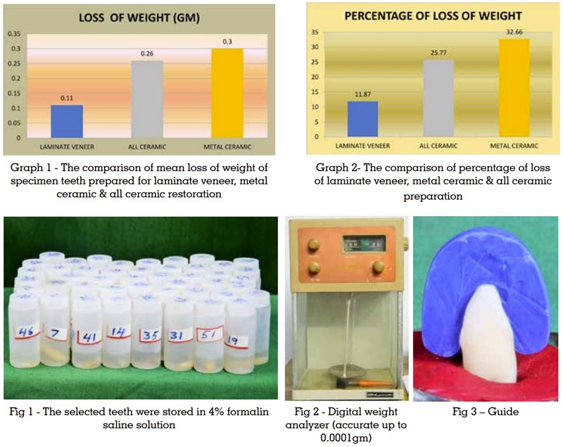
The selected teeth were stored in 4% formalin
saline solution for four weeks. (Fig – 1) Teeth were
made free from stain, calculus and soft tissue, by
using an ultrasonic scaler, polishing brush, and
pumice water mixture. They were then examined
under microscope at 2.5× magnification to ensure
that they are free from fracture, caries, restoration,
crazing.5
The bucco-lingual and mesio-distal dimensions
of each specimen was measured at the cemento-enamel junction by using a thickness gauge.
The baseline mass for each tooth was measured with the help of a digital analyzer (accurate up to
0.0001gm) and recorded at the beginning of the
study. (Fig-2) All teeth were blotted for 10 minutes
on absorbent paper towel prior to weighing.6
The
teeth were then mounted on a mounting jig prepared with impression compound prior to preparation. The specimen teeth were then prepared for
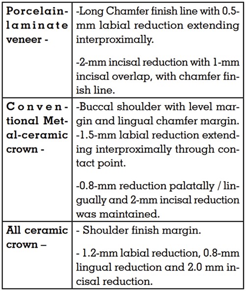
receiving Porcelain laminate veneer, Conventional
metal ceramic and All-ceramic restoration according to pre-determined standardized preparation design. The pre-determined standardized
preparation designs followed in this study were
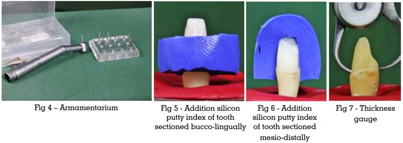
as following using proper armamentarium (Fig-3
and Fig-4) – 6,7
Two addition silicon putty indexes were made
of each tooth and sectioned bucco-lingually
and mesio-distally and used as reference guide
throughout the reduction procedure to standardize
and estimate the amount of tooth reduction (Fig-5
and Fig-6). This was achieved by measuring the
distance between the tooth and the fitting surface
of the reduction index.8
(Fig-7)
After preparation each specimen tooth was kept
in 4% formalin-saline solution and then blotted in
absorbent paper towel for 10 minutes. The teeth
were then weighed in a digital analyzer.
The weight was calculated as follows – 9
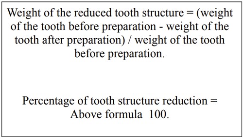
For statistical analysis, descriptive statistics were
used to analyze the percentage of tooth mass reduction. Student’s t-test was used to compare the
mean tooth reduction among the different types of
preparation, and a p value <0.05 was considered
statistically significant.
On calculation of final weight difference between
laminate veneer & all ceramic, laminate veneer &
metal ceramic and all ceramic & metal ceramic
using unpaired t test, it was noticed that there is
no statistically significant difference in mean final
weight between Laminate Veneer & All Ceramic,
and All Ceramic & Metal Ceramic. (P value >0.05).
While, there was statistically significant difference
in mean final weight between laminate veneer &
metal ceramic. (p value <0.05). (Table-1)
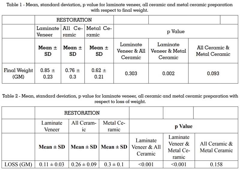
The loss of weight between laminate veneer and all
ceramic, laminate veneer and metal ceramic and
all ceramic and metal ceramic by using unpaired
t test, no statistically significant difference was
seen in mean loss of weight between all ceramic
& metal ceramic. (p value >0.05). Whereas, there
was statistically significant difference in mean loss of weight between laminate veneer & all ceramic
and laminate veneer & metal ceramic. (p value
<0.05). In laminate veneer there was significantly
less loss of weight as compared to the other types
of preparations. (Table -2)
The mean weight loss for porcelain laminate veneer was (0.11 ± 0.03) gm, for all ceramic crown
preparation is (0.26 ± 0.09) gm, for metal ceramic
it is (0.30 ± 0.10) gm. There was no statistically
significant difference in mean weight loss between
all ceramic and metal ceramic but there was statistically significant difference in mean loss in weight
between laminate veneer and metal ceramic, in
laminate veneer there was statistically significant
difference in mean loss of weight as compared to
other types of preparation. (Graph – 1)
The percentage of loss of tooth structure between
laminate veneer and all ceramic, laminate veneer and metal ceramic, all ceramic and metal
ceramic were statistically significant. (p value
<0.05). In laminate veneer percentage of weight
loss is minimum followed by all ceramic & metal
ceramic. (Table – 3)
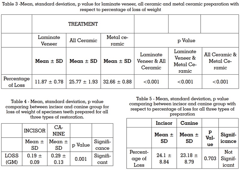
The percentage of loss of tooth structure between
laminate veneer and all ceramic, laminate veneer
and metal ceramic, all ceramic and metal ceramic
were statistically significant. In laminate veneer
preparation percentage of weight loss was minimum followed by all ceramic and metal ceramic.
The mean percentage of weight loss obtained for
laminate veneer as (11.87 ± 0.78), for all ceramic
(25.77 ± 1.93) and for metal ceramic (32.66 ±
0.88). (Graph – 2)
On comparison of incisor group of teeth with canine group after tooth preparation for all selected
types of restorations, the mean loss in weight was
significantly higher for canine group as compared
to incisor group. (p value <0.05). (Table 4)
The percentage of loss of tooth structure for incisor and canine group for all selected types of
restorations were not significantly difference. (p
value >0.05). (Table 5)
The incisor group of teeth when compared with
canine group after tooth preparation for all selected types of restorations, the mean loss of weight
for incisor group is (0.19 ± 0.09) gm and for the
canine group it was (0.29 ± 0.13) gm, which was
statistically significant. So, it can be said that in
canine group loss of tooth structure is more when
compared to incisor group. The percentage of loss of tooth structure for incisor and canine group for
all selected types of restorations was (24.1 ± 8.84)
and (23.18 ± 8.79) respectively, there was no significant difference in mean percentage of weight
loss across canine and incisor group.
The importance of quantification of tooth structure
removal cannot be over emphasized. There is very
limited number of studies that quantified the tooth
structure loss with respect to different preparation
designs. Different methods have been described
to measure the amount of tooth structure removal
associated with different preparation designs.
[9,10] Given the accuracy, ease and simplicity
gravimetric analysis was employed to measure
the tooth structure removal.
Commonly practiced treatment options for anterior
tooth restoration are porcelain laminate veneer,
all ceramic full veneer crown and conventional
metal ceramic crown.10,11 So, in this study, all three
forementioned preparations were chosen. Sixty
anterior teeth were chosen as sample and underwent preparation for porcelain laminate veneer,
all ceramic full veneer crown and metal ceramic
crown.
For anatomical crown, Edelhoff and Sorensone
had quantified and compared the tooth weight
only of acrylic resin typodont teeth with different
preparation designs. The authors had reported
that different preparations designs resulted in significant differences in the amount of tooth removed.5,10 – 13
Based on the findings of Hussain Sela .K F , McDonald Aibhe and Moles David R a baseline
could be established for comparison in the current
study. They found the marginal mean percentage
of tooth structure loss at the end of preparation
for porcelain laminate veneer to be 80.7% and for
metal ceramic preparation it was 61.30%, which
were similar to the findings of our study.8
The results in case of loss of tooth structure in the
canine group being more compared to incisor
group loss was in contradiction to Hussain Sela K
F , McDonald Aibhe and Moles DR’s study where they found that incisor group of tooth preparation
demanded more tooth structure removal than
that of canine group.8
This phenomenon could be
explained by the use of heterogonous morphology
of collected sample of tooth. There might have
also been a racial variation in tooth morphology
particularly in anterior teeth.
It should be noted that in this ideal preparation
design only specific requirement of the material
were considered as a factor for tooth structure
removal beyond that, other criteria might control
the preparation design in the patient’s mouth.
By conducting this experiment on similarly sized
single rooted natural teeth and reduction by a single operator, it was attempted to minimize both the
amount of morphological and operator variability
encountered and subsequently it ensured that the
results obtain were as accurate as possible with
regards to change in mass. Further investigations
are needed to confirm the relative contribution of
the loss of tooth structure with respect to different
preparation design.
Limitations –
In this study only two preparation designs i.e.
partial and complete coverage were used for the
tooth morphology. Many more designs could have
been incorporated. The sample size of the tooth
studied were not equal and newer methods for quantifying removed tooth structure could have
been used.
Future prospects –
The sample size can be increased to achieve more
definite results. The number of designs of tooth
preparation can be increased to incorporate more
variability. In this study manual techniques were
used, with the advent of technology, digitilisation
of the entire process can be done using digital
scans, computer added designing and computer
aided milling.
Within the limitations of this in vitro study the following conclusions can be drawn –
Though the metal ceramic restorative procedure
is most widely practiced across the world it demands highest amount of tooth structure removal.
So, when clinical condition permits, considering
this restorative procedure, the clinician may think
over the other modalities of treatment once more.
Minimally invasive veneer preparation offers a
tremendous advantage over all ceramic crown and
conventional metal ceramic crown preparation.