Purpose: To evaluate whether the incorporation of graphene nanoparticles to PMMA improves its thermal conductivity properties. Materials and Methods: In this in vitro experiment, samples of 10 PMMA acrylic disks, 5 disks with graphene nanoparticles (test group) and 5 disks without graphene nanoparticles (control group) were fabricated and subjected to thermal conductivity testing. The sample disks were placed between the two plates of the instrument. The results were analyzed using the Mann Whitney U- test (p<0.05). Results: PMMA disks without graphene additives showed superior thermal conductivity than the test group (p= 0.028). Conclusion: Under the given conditions, acrylic disks with graphene additives did not conduct heat more than the disks without graphene additives. Sographene did not significantly improve the thermal properties of PMMA.
Key words: graphene in dentistry, thermal conductivity, polymethyl methacrylate
Various biomaterials are used in Prosthodontics
to fabricate removable prostheses for completely
and partially edentulous patients. One of the
most commonly used materials with decades of
evidence is polymethyl methacrylate (PMMA).
Polymethyl methacrylate has stood the test of time
as a denture base material and holds its efficacy
as a satisfactory denture base material till date.
It was introduced in the year 1937 by Dr. Walter
Wright and has its pros and cons1.
PMMA has been employed successfully as a
denture base material owing to its light weight,
ease of fabrication, low cost, acceptable aesthetics,
biocompatibility, low water sorption, and
adequate strength; at the same time, it has certain
disadvantages such as low fatigue resistance,
low coefficient of elasticity and low thermal
conductivity. Among these, of prime concern is the
low thermal conductivity of PMMA which reduces
the gustatory response and palatability of food
for the patient2. Improved thermal conductivity
of polymethyl methacrylates would be a boon to
removable denture wearers thereby resulting in
enhanced the acceptance of these dentures.
“Thermal conductivity, of a substance, is defined as the quantity of heat in calories, or Joules, per
second, passing through a body 1 cm thick with a
cross section of 1 cm square when the temperature
difference is 1-degree Celsius”. The value of
thermal conductivity for PMMA is 0.2 W/m/degree
Celsius3.
To overcome this shortcoming, various materials
have been used to improve the thermal conductivity
of PMMA. Metal oxides like aluminium oxide,
titanium oxide, zirconium oxide, silicon carbide
filler powders, hydroxyapatite ceramic powders,
silver nanoparticles, and carbon particles have
been incorporated and have improved the thermal
conductivity without affecting the strength of the
acrylic denture base. Also, metal denture bases
have been used by many clinicians to overcome
this disadvantage1,4,5.
The present study was conducted to test if the
addition of a material, named Graphene, improved
the thermal properties of dental polymers or not.
Graphene which is a basic construction of graphite
consists of carbon atoms arranged in a honey-comb
structure in the form of flat thick sheets. It has broad
potential applications in biomedical engineering
and biotechnology, such as in DNA detection, drug delivery, cancer therapy, etc. Owing to its various
excellent mechanical, thermal, electrical and antimicrobial
properties6,7,8 graphene is now being used
in dentistry for detection of bacteria in dental caries
and periodontal disease, in restorative dentistry
for strong dental fillings that do not corrode in the
oral cavity, as fillers in dental adhesives to prevent
secondary caries, in bone tissue engineering, in
periodontal tissue regeneration, in cancer therapy,
as coating on implants to improve osseointegration
and lastly its antibacterial properties which kills
bacterial cells through cell- wrapping and celltrapping
mechanism. Due to these enhanced
capabilities, graphene is being incorporated in
dental biomaterials such as metals, ceramics,
and polymers. Different graphene derivatives can
achieve good dispersion within different polymers
and can be easily processed8.
Previous studies have proved that the addition of
graphene to PMMA does not affect the impact and
flexural strength. They are also biocompatible and
nontoxic in the oral cavity. Graphene has been
proved to be a superior thermal conductor in other
biomedical applications. But this valuable property
has not been investigated in dentistry so far6.
Swami P, Sanyal P, Guru R, Kore A9 evaluated and compared the addition of graphene and carbon
fillers on the flexural and impact strength of PMMA.
They concluded that 0.5% by weight of carbon
fillers increased the flexural and impact strength
of PMMA followed by graphene combined with
0.25% carbon and 0.25% graphene nanofillers.
Guazzo R10 reviewed the uses of graphene
nanomaterials in dentistry. They discussed the
interaction between graphene nanomaterials with
immune system cells and antibacterial activity of
graphene nanomaterials. They concluded that
further studies are required to evaluate the possible
long term toxicity and their derivatives in the oral
cavity. Malik S11 described a new,easy and low-cost
method to make large amount of FLG few layered
graphene with multi-layered graphene and its
inclusion into dental polymers. It was concluded
that the new few layered graphene material
incorporated in the dental polymer enhanced
the physicomechanical properties as there was
27% increase in mean compressive strength and
mean compressive modulus increased by 22%.
Shradhanjali A, Bouzid T, Sinitskii A, Lim J12
and Kulshrestha S et al7 reviewed the use of
graphene for dental implant applications.
Osseointegration of dental implants has been proven to improve after coating the implant surface
with graphene oxide and reduced graphene oxide.
The osteogenic differentiation of mesenchymal
stem cells significantly improved on the implant
surface and also increased the antibacterial
properties. But further studies have to be done
to reveal how graphene-based implant coatings
would produce better implant actions. Lakshmi
K, Rao GU, Arthiseethalakshmi S, Mohamed
MSK13 reviewed the properties, manufacturing
methods, applications of graphene in general
and biomedical fields and also in dentistry.
In dentistry graphene is used for detection of
bacteria in dental caries and periodontal disease,
in restorative dentistry for strong dental fillings
that do not corrode in the oral cavity, as fillers in
dental adhesives to prevent secondary caries,
in bone tissue engineering, in periodontal tissue
regeneration, in cancer therapy, as coating
on implants to improve osseointegration and
antibacterial properties. Thus, they concluded that
further research is needed for its use in dentistry.
The aim of this study is to investigate whether
the addition of graphene nanoparticles in PMMA
improves the thermal conductivity. Null hypothesis
of this study is that the addition of graphene nanoparticles in PMMA does not affect the thermal
conductivity.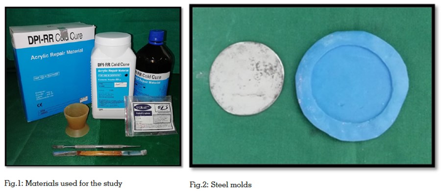
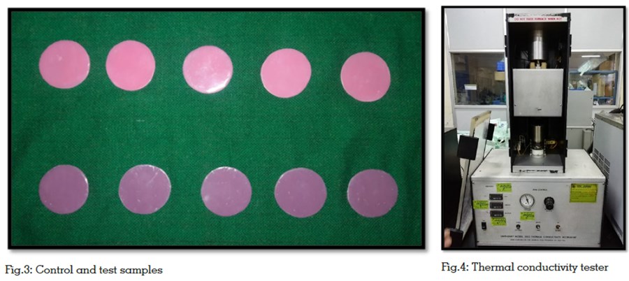
Two stainless steel disks were constructed by
cutting solid stainless steel plates into the desired
shape and dimension of 50 x 2±0.5mm (Fig.2)14.
Ten putty molds, (Virtual, Putty Base Regular Set,
Ivoclar Vivadent, Italy) five from each disk were
fabricated. From these molds, ten acrylic (DPI-RR
Cold Cure, Dental products of India, Mumbai)
specimens were made, five without graphene
additives to serve as a control group and five with
graphene additives (UGRAY, United Graphene,
United Nanotech Innovations Pvt. Ltd, Bangalore)
to serve as a test group (Fig.3).
An electronic balance (Shimadzu, India) was used
to measure the graphene and polymer powders.
2% by weight of graphene (particle size of 25μm)
was added in 100g of polymer: 33ml of monomer
for the five test samples9. Similarly, the control
samples were prepared using the same polymer:
monomer ratio without the addition of graphene.
The acrylic mixture was then poured into the putty
molds and polymerized in a pressure pot (Vertex,
curing pressure pot, Multicure) at 10MPa for 30
minutes. The polymerized disks were then finished and polished using tungsten carbide trimmers
and silicone burs.
The samples thus obtained were tested using a
thermal conductivity instrument (Unitherm Model
2022, Pittsburgh, USA)(Fig.4). This instrument uses
the guarded hot plate principle to conduct heat15.
Two plates, namely a hot plate and a cooling
plate are in built in this instrument. The sample
material was set between the two plates. One
plate was heated and the other plate was cooled.
The temperature of the plates were observed until
they were constant (mean temperature of 55℃
was maintained). The steady-state temperatures,
the thickness of the sample and the heat input
to the hot plate were used to calculate thermal
conductivity of the samples.
The results thus obtained were subjected to
statistical analysis using the Mann Whitney Utest,
a non-parametric test which considers the
magnitude of the differences via ranks to test if
two samples come from identical populations.
The values obtained for the control group without
the addition of graphene ranges from 0.123 W/m/
ºC to 0.135 W/m/ºC with a mean of 0.129 and a
standard deviation of 0.004(Table 1). The values
for the test group ranges from 0.115 W/m/ºC to 0.128 W/m/ºC with a mean of 0.112 and standard
deviation of 0.012 (Table 1). The p-value was
found to be 0.028. This shows that the values
have decreased consistently for the test group
when compared to control group. The p-value is
less than 0.05 which proves that the results are
statistically significant.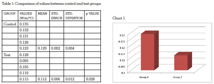
Polymethyl methacrylate has been dominating
the dental industry for many years due to its near
ideal mechanical and physical qualities. Numerous
studies have been conducted in the past to enhance
these properties by incorporating various fillers
and fibers16,18. The enhancement of the thermal
conductivity of PMMA has been experimented
using materials like aluminium oxide, titanium
oxide, zirconium oxide, silicon carbide filler
powders, hydroxyapatite ceramic powders, silver
nanoparticles and carbon particles17,19.
In the present study, ten acrylic disks measuring 50
x 2±0.5mm were fabricated for thermal conductivity
testing, five with graphene additives and five
without graphene additives. These disks were
polymerized and subjected to thermal conductivity
testing. The values thus obtained were subjected
to statistical investigation using Mann Whitney
U non-parametric test. The p-value was found to
be 0.028 (p<0.05). This shows that the study is
statistically significant and the null hypothesis
is rejected.
Results prove that thermal conductivity had
reduced for the test group when compared to the
control group. Thus, the addition of graphene
additives has reduced the thermal conductivity
of PMMA. The possible reasons for this finding
could be the proportion of graphene in polymer/
monomer was inadequate. The quantity of
graphene incorporated in the test samples lead
to an acceptable discoloration which can be used
for characterizing dentures. But the addition of more amount of graphene led to an unaesthetic
dark discoloration of the material which would be
unsuitable for fabricating the prosthesis9. Also,
there could have been agglomeration of graphene
particles since they were not subjected to a process
called as ultra probe sonification9. The thermal
conductivity instrument (Unitherm 2022, ASTM E
1530, CIPET/PTC/027. Anter Corporation, USA) can
conduct heat equally only if the sample thickness
is constant throughout with a variation of ± 0.5mm.
There could have been possible variations in
the thickness of the sample while finishing and
polishing,this could have lead to varying values.
The study was conducted involving a small sample
size, further investigations can be done using a
larger sample size. Also, heat cure denture base
can be used.
Within the limitations of the present study, it can be concluded that,