The socket shield procedure is a new technique in aiding the preservation of the vulnerable buccal bundle bone during immediate implantation . Latest digital dentistry techniques are used to assist in performing a complex procedure
Key words: socket shield, digital dentistry
The extraction of teeth especially in the aesthetic zone, invariably produces changes that can adversely affect the buccal soft tissue and the underlying buccal bone. This can result in a compromise in both aesthetics and function leading to unhappy patients and sub optimal results especially during immediate implant placement.
The conventional way is to use biomaterials soft tissue grafting and guided tissue regeneration and guided bone regeneration. However these techniques increased the complexity and cost of the procedure.
A novel technique was first described by Hurzeler et al where the buccal bone and soft tissue was preserved by retaining the buccal fragment of the root to be extracted ( Hürzeler, Zuhr et al. 2010, Baumer, Zuhr et al. 2015)
A female patient with a high smile line came to the office with a failing post retained crown on the UR4,( Fig 1 )this patient had previous orthodontic treatment and hence are missing the second premolars as well. So the visibility of the UR4 is quite obvious. She had good oral hygiene and well motivated to keep the smile as intact as possible. She also had a thin biotype. The patient was not keen on gingival grafts and biomaterials and had a limited budget as well.
After further discussion, the socket shield technique was demonstrated and consent gained.
The old root filling was removed completely and radiopaque CaOH was placed for one week in the root canals. CBCT was performed and a surgical guide was printed from the CT data to help with precise cut of the root (Fig 2). The radiopaque CaOH helped in precisely measuring the root for the socket shield.
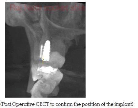
Tungsten carbide burs with a long shank on a speed increasing electric handpiece was used to section the root using the surgical guide under an operating microscope. Osteotomy was performed conventionally palatal to the buccal fragment. Note the radiopaque CaOH in the buccal fragment of the root. This started out as the classical Socket Shield but ended up as a modified root submergence technique. A 3.8x11mm Sweden and Martina implant system was used. Post op CBCT taken shows the implant in the desired position palatal to the buccal fragment and also not touching the root fragment (Fig 3)
The socket shield techniques are experimental, there are no long term studies other than case reports in support of this technique eventhough it was first described in 2010. It is very technique sensitive and should not be attempted by a beginner in implantology. Turbines should not be used for this technique as the carbide bur can judder in a turbine and the precision and controlled torque can be compromised. High magnification and co axial illumination is recommended.
Digital technology assists in making a complex procedure predictable however this is not a technique for beginners. If the fragment gets mobile , conventional bone and soft tissue surgical skills and GBR has to be in place . This is an extra tool in the armamentarium of an experienced implant surgeon and Prosthodontist.