Maxillary defects resulting from surgical resection, trauma, or congenital abnormalities can significantly impair speech, mastication, swallowing, and esthetics. Conventional obturator fabrication techniques are often complex, time-consuming, and highly technique-sensitive. The advent of digital dentistry has introduced innovative approaches involving intraoral scanning, computed tomography (CT), computer-aided design/computer-aided manufacturing (CAD/CAM), and three-dimensional (3D) printing technologies. These advancements have enabled clinicians to improve treatment efficiency, accuracy, and patient comfort. This review summarizes the current evidence regarding digital workflows in obturator fabrication and discusses their advantages, limitations, and future prospects.
Key words: Obturator, CAD/CAM, Digital Dentistry, 3D Printing, Maxillofacial Prosthodontics
Maxillary defects create communication
between the oral and nasal cavities, resulting in
compromised speech, swallowing, mastication,
and facial esthetics. Obturator prostheses
remain the most commonly used method for rehabilitation of these patients. Conventional
techniques involve multiple clinical and
laboratory procedures and are often associated
with patient discomfort and inaccuracies1.
Recent advances in digital dentistry have
transformed maxillofacial prosthodontics by
enabling virtual treatment planning, digital
impression making, CAD-based prosthesis
design, and additive manufacturing techniques2.
Studies have demonstrated that digital
workflows can reduce treatment time, improve
patient comfort, and enhance reproducibility of
prostheses3.
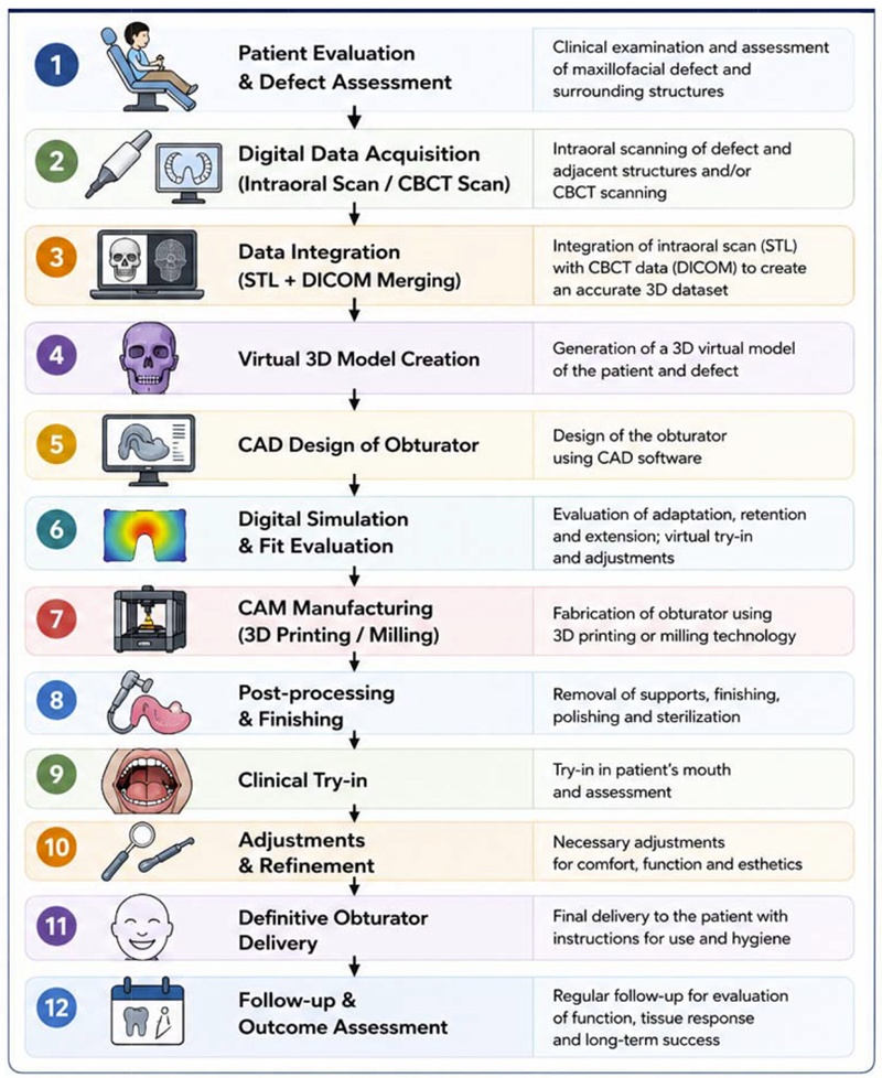
The first step in digital obturator fabrication is
acquisition of anatomical information through
intraoral scanners, CT, CBCT, and facial
scanning systems. Digital impressions eliminate
many shortcomings associated with conventional
impression materials and techniques4.
Tasopoulos et al. described the use of intraoral
scanning combined with postoperative CT data
to fabricate a two-piece hollow bulb obturator.
The authors reported improved patient comfort
and satisfactory clinical accuracy while avoiding
conventional impression procedures5.
Digital scanning is particularly beneficial in
patients with severe trismus, extensive defects,
or limited mouth opening, where conventional
impression making is challenging6.
Following data acquisition, the digital files are
imported into CAD software for virtual design of
the prosthesis. CAD software allows visualization
of undercuts, defect morphology, and framework
design before manufacturing7.
Virtual planning facilitates modifications without
repeating clinical procedures and improves
communication among prosthodontists,
surgeons, and laboratory technicians2.
Three-dimensional printing has emerged as a
major advancement in obturator fabrication.
Additive manufacturing enables fabrication
of lightweight hollow obturators with complex
geometries that are difficult to achieve using
conventional laboratory techniques8.
Koyama et al. presented a workflow that
integrated optical scanning and digital molding
technologies to fabricate a single-piece hollow
obturator, simplifying laboratory procedures
and minimizing fabrication errors9.
Recent reports have demonstrated successful
fabrication of obturators using resin-based
printers,
selective laser sintering, and
polyetheretherketone (PEEK) frameworks5.
CAD/CAM technology enables both additive and
subtractive manufacturing approaches. Milled
prostheses offer improved material homogeneity
and mechanical properties, whereas printed
prostheses provide greater flexibility in design7.
Digital and cast-free workflows have recently been introduced, allowing fabrication of
definitive hollow obturators without conventional
stone casts. Such approaches reduce laboratory
errors and improve overall treatment efficiency10.
Digital technology has also facilitated the
fabrication of surgical obturators. Through
virtual surgical planning and preoperative
digital design, prostheses can be fabricated
before surgery and delivered immediately
following maxillectomy procedures11.
This approach minimizes rehabilitation time and
improves postoperative patient management.
The major advantages reported in the literature include1,3:
Systematic reviews have concluded that digital workflows significantly reduce working time while enhancing patient acceptance and satisfaction11.
Despite promising results, several limitations remain1,3:
Most available evidence consists of case reports and technical reports, highlighting the need for randomized clinical studies.
Future developments may include artificial
intelligence-assisted design, automated CAD
workflows, advanced printable biomaterials, and
integration of augmented reality technologies.12
Digital workflows are expected to become
increasingly accessible and may eventually
become the standard approach for maxillofacial
prosthetic rehabilitation.
Digital workflows have significantly transformed
obturator fabrication. The combination of
digital scanning, CAD design, and additive
manufacturing offers improved accuracy,
efficiency, patient comfort, and reproducibility
compared with conventional methods. Although
challenges remain, current evidence suggests
that digital technologies represent a promising
future direction for maxillofacial prosthodontics.