Aim and background: Prosthodontic rehabilitation of
completely edentulous patients with maxillary defects
presents significant challenges due to the absence
of natural teeth for retention and support. Successful
obturator therapy requires careful consideration of
defect size, available tissue undercuts, and prosthesis
weight. Hollow bulb obturators are often indicated
to reduce prosthesis weight and improve retention;
however, the choice of fabrication technique should
be individualized. This case series aims to illustrate
the clinical application of three different obturator
processing techniques in completely edentulous
maxillectomy patients and to highlight factors
influencing technique selection.
Case description: Three completely edentulous
patients with maxillary defects following surgical
resection were rehabilitated with definitive obturator
prostheses. In the first case, a conventional solid
closed-bulb obturator was fabricated due to
the relatively small defect size and acceptable
anticipated prosthesis weight. In the second case,
a hollow bulb obturator was processed using the
lost-wax bolus technique to reduce the weight
associated with a larger unilateral defect. In the third case, involving a bilateral total maxillectomy
defect with limited mouth opening and absence of
hard tissue support, a hollow bulb obturator was
fabricated using the lost salt technique to achieve
maximal weight reduction. Clinical outcomes were
evaluated based on prosthesis retention, speech
intelligibility, swallowing function, aesthetics, and
patient satisfaction.
Conclusion: All three patients demonstrated
satisfactory functional and aesthetic outcomes
following prosthodontic rehabilitation. The selection
of obturator processing technique should be guided
by defect extent, available retention, and the need for
weight reduction. Individualized treatment planning
contributes significantly to successful rehabilitation
in completely edentulous maxillectomy patients.
Clinical significance: This case series provides
clinical options for selecting obturator fabrication
techniques based on defect characteristics and
prosthetic requirements. Appropriate technique
selection can improve retention, reduce prosthesis
weight, and enhance functional outcomes in
edentulous patients with maxillary defects.
Key words: Palatal obturators, mouth, edentulous, neoplasm, prosthodontics
Maxillary defects arising from surgical resection
of neoplasms result in oronasal communication,
causing profound impairment of speech,
deglutition, and facial aesthetics. Prosthodontic
rehabilitation with an obturator prosthesis
remains the mainstay of management, offering
restoration of oronasal separation and functional
recovery without the morbidity of reconstructive
surgery.1 The challenge of obturator fabrication
is compounded in completely edentulous
patients. Retention must be derived solely from
tissue undercuts, scar bands, and the peripheral
seal over residual alveolar ridges.2,3 Prosthesis
weight is a critical variable in obturator design.
A large solid obturator may be excessively heavy,
overwhelming available soft tissue retention
and causing patient discomfort. Hollowing the
obturator bulb is therefore frequently necessary.
Several techniques for creating a hollow
bulb have been described, each with distinct
procedural and clinical characteristics.4,5
This case series presents three completely
edentulous maxillectomy patients rehabilitated
with definitive obturators, each fabricated using
a different processing technique, illustrating
how defect extent, available retention, and tissue
status guide technique selection.
A 58-year-old male patient presented with a
history of squamous cell carcinoma of the left
maxilla managed by partial maxillectomy
approximately 14 years ago. He was completely
edentulous in the maxilla and presented with a
left-sided hard palate defect extending across
the midline with an intact soft palate. (Figure 1a)
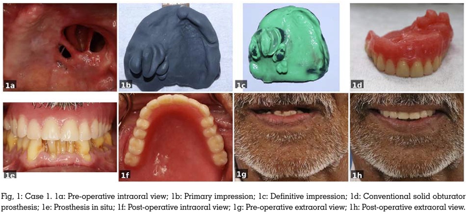
A primary impression was made using impression
compound to record the defect and the residual
ridge. (Figure 1b) A custom tray was constructed,
border moulding was performed with low-fusing
compound, and the definitive impression was
made using light-body addition silicone. (Figure
1c) Jaw relation records were obtained using a
record base and occlusal rims. Conventional complete denture teeth were arranged, and a
wax try-in was conducted to verify aesthetics
and occlusion.
Given the modest defect dimensions and limited
vertical depth, the anticipated prosthetic weight
was within acceptable limits. A conventional solid
closed-bulb obturator was therefore processed
using heat-polymerised polymethylmethacrylate
(PMMA) without hollowing. (Figure 1d) Post
insertion adjustments were completed over
subsequent
reviewappointments.
Residual
alveolar ridge, intact soft palate and scar tissue
along the lateral defect margins provided
palpable undercuts that were engaged for
retention. (Figure 1e and Figure 1f). At the
one-year follow-up, the patient demonstrated
marked improvement in speech intelligibility
and deglutition. Adequate prosthetic stability
was maintained, and the patient expressed
satisfaction with functional and aesthetic
outcomes. (Figure 1g and Figure 1h)
A 66-year-old male patient presented with a
maxillary defect following partial maxillectomy for
acanthomatousameloblastoma of the
right maxilla. Clinical examination showed a
completely edentulous maxilla withthe defect
extending to the right side, crossing the midline,
making it substantially larger than in Case 1.
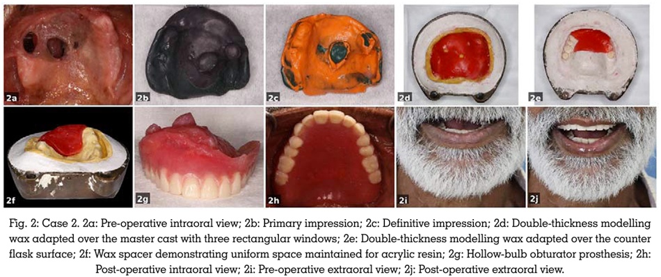
(Figure 2a) Clinical procedures mirrored those
of Case 1. (Figure 2b and Figure 2c)
In view of the larger defect volume, a solid
obturator was expected to produce excessive
prosthetic weight that would overwhelm the
available retention. Hollowing was planned
using the lost-wax bolus technique. Following
investing and dewaxing, double-thickness
modelling wax was adapted over the master cast
and counter flask surfaces to create a uniform
spacer, with three rectangular windows to
ensure consistent acrylic wall thickness. (Figure
2d and Figure 2e) Following a second dewaxing
cycle and PMMA packing, the wax bolus was
repositioned as a spacer and the prosthesis was
processed conventionally. (Figure 2f) 3 holes were
made on the cameo surface and the obturator
was flushed with hot water to eliminate the wax,
followed by sealing using auto-curing resin. The
bilateral tuberosities provided primary hard tissue retention, supplemented by engagement
of scar bands at the lateral margin of the defect.
(Figure 2g and Figure 2h) Speech, swallowing,
and aesthetics improved at the six-month follow
up. (Figure 2i and Figure 2j)
A 68-year-old male patient reported with
a bilateral maxillary defect following total
maxillectomy for squamous cell carcinoma. He
had received radiation therapy a decade prior
to presentation and was completely edentulous.
The defect was the largest of the three cases,
with complete absence of residual hard palatal
tissue and no bony undercuts available. The
patient had reduced mouth opening secondary
to post-surgical and post-radiation fibrosis.
He had previously worn an obturator without
denture teeth and wished to continue with this
preference.
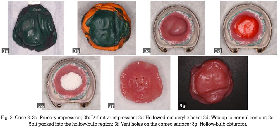
Primary and secondary impressions were made
in a conventional manner. (Figure 3a and Figure 3b) Given the total maxillectomy defect volume,
a solid obturator would have been excessively
heavy, compromising retention. The lost salt
technique was therefore selected. Processing was
accomplished using base-flask with 2 counter
lids. Initially, a hollowed-out base was acrylized.
(Figure 3c) Next, the lid of the obturator wax
waxed-up and invested. (Figure 3d) Following
dewaxing, finely granulated salt was packed
into the hollow bulb region to a uniform depth.
(Figure 3e) The assembly was flasked and
processed conventionally. Following deflasking,
3 holes were made on the cameo surface and
the prosthesis was immersed in warm water,
allowing complete dissolution and evacuation
of the salt crystals yielding a smooth, uniformly
hollow chamber. (Figure 3f) The holes were then
sealed using auto-curing acrylic resin. Retention
was primarily achieved through posterior scar
band engagement and soft tissue undercuts at
the lateral margins of the defect. (Figure 3g). At
the six-month follow-up, speech and swallowing
showed meaningful improvement. The markedly reduced prosthetic weight was critical to
achieving functional retention in the complete
absence of hard tissue support.
The three cases presented demonstrate the
spectrum of clinical complexity in completely
edentulous
obturator
rehabilitation and
illustrate how processing technique selection
must be individualised to patient-specific
factors. Unlike dentate patients, the completely
edentulous maxillectomy patient has no teeth
to serve as retentive abutments. The prosthesis
must rely entirely on tissue undercuts, peripheral
seal, and scar band engagement. Devlin and
Barker underscored the particular difficulty of
achieving adequate retention in the edentulous
maxillectomy patient, noting that the reduction
in denture-bearing area combined with altered
ridge morphology demands careful impression
technique and prosthesis design.3 Yenisey et
al. similarly emphasised the use of anatomical
undercuts within and around the defect as the
primary retentive mechanism.2
Prosthesis weight emerges as a dominant factor
in these patients. A heavy prosthesis exerts a
displacing force that soft tissue retention alone
cannot resist, compromising stability and
patient comfort. Hollowing the obturator bulb
substantially reduces this load and has been
shown to improve retention and patient-reported
outcomes.4,6
The conventional solid technique employed in
Case 1 was appropriate for a smaller defect
with limited vertical extension. In this context,
the simplicity and structural durability of a
solid prosthesis outweigh the modest weight
disadvantage2. The wax bolus technique
offered an efficient solution for a larger defect
requiring weight reduction. Its limitation lies in the requirement for predictable wax adaptation
within the bulb, best suited to defects of moderate
and regular geometry. Nimonkar et al. reviewed
various hollow bulb methods and noted that wax
based spacer techniques provide acceptable
outcomes in moderately sized defects when wall
thickness can be controlled.4
The lost salt technique provided the greatest
degree of hollowing for the most complex defect.
Salt crystals pack uniformly into cavities of any
shape, enabling reliable hollowing of irregularly
contoured bulbs where wax adaptation would
be imprecise.5,7 Complete salt dissolution leaves
no residue, and the smooth internal chamber
ensures optimal weight reduction. The altered
prosthodontic approach required for a bilateral
total maxillectomy including limited mouth
opening, dependence on soft tissue alone,
is consistent with the challenges reported by
Cheng et al.8
Prosthodontic rehabilitation of completely
edentulous patients following maxillary resection
is a challenging undertaking, demanding careful
assessment of defect extent, available retention,
and individual patient factors. This case series
demonstrates that, with meticulous clinical
planning and technique individualisation,
satisfactory outcomes in speech, deglutition,
and aesthetics can be achieved even in the most
complex cases.