Introduction: Maxillary tumor resection compromises
the retention, stability, and support necessary for
conventional dentures. Adjunctive radiotherapy
exacerbates mucosal friability and causes xerostomia,
making heavy prostheses clinically problematic.
Case Presentation: A 71-year-old male presented with
edentulous arches following an anterior maxillectomy
and radiotherapy for squamous cell carcinoma.
Clinical examination revealed total loss of the anterior
maxilla, right tuberosity hypertrophy, a hypermobile
mandibular anterior ridge, and advanced bilateral
posterior resorption. To optimize retention and
minimize mucosal loading, a lightweight hollow
maxillary complete denture was fabricated using
a customized soap-spacer technique, paired with
a conventional mandibular denture.
Conclusion: The hollow maxillary denture
successfully compensated for missing bone support without overloading the irradiated mucosa. This
cost-effective protocol provided a highly functional
prosthesis, presenting a viable pathway for post
oncological prosthodontic rehabilitation.
Key words: maxillectomy, radiotherapy, hollow denture, oral rehabilitation
Surgical maxillectomy for oral malignancies
severely reduces the mechanical supporting
tissue area required for complete dentures,
while adjunctive radiotherapy compromises
mucosal micro-vascularity and causes chronic
xerostomia.1,2 The resulting loss of the peripheral
seal renders heavy, conventional acrylic
resin dentures highly prone to gravitational dislodgement and recurrent mucosal trauma.3
Fabricating
a
hollow maxillary denture
significantly reduces the total prosthetic mass,
thereby lessening gravitational dislodging
forces and preserving compromised underlying
structures.4 While advanced digital workflows
offer precise modern alternatives, traditional
mechanical hollowing techniques remain vital
for accessible, cost-effective care.5 This case
report outlines a predictable, low-cost laboratory
modification using a custom soap spacer to successfully rehabilitate a post-maxillectomy
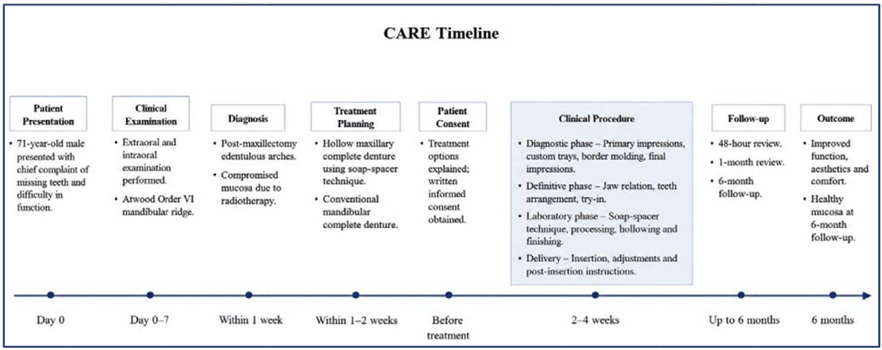
edentulous patient as shown in the below CARE
timeline.
A 71-year-old male patient presented to the
Department of Prosthodontics seeking prosthetic
rehabilitation for missing teeth. His medical
history was significant for type 2 diabetes
mellitus, which was well-controlled with oral
hypoglycemic agents. The patient had been diagnosed with poorly differentiated squamous
cell carcinoma of the maxilla two years prior.
His oncological treatment plan consisted of a
surgical anterior maxillectomy (premaxillary
region) followed by external beam radiation
therapy (total dose: 60 Gy), completed 18 months
before presentation.
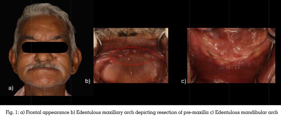
Extraoral examination revealed a significant
loss of lip support in the subnasal region [Figure 1a]. Intraoral examination revealed completely
edentulous maxillary and mandibular arches.
In the maxillary arch, the alveolar ridge in the
premaxillary region was completely absent due
to surgery; however, mucosal continuity remained
intact without open oronasal communication.
Hypertrophy of the right maxillary tuberosity
was noted. The mandibular arch exhibited a
hypermobile, flabby anterior residual ridge
and advanced residual ridge resorption in the bilateral posterior segments, classified as
Atwood’s Order VI [Figure 1b, 1c].
Given the anatomical constraints and compro mised mucosal support, the below prosthetic workflow was initiated:
Following clinical try-in, the trial maxillary
denture was invested in a dental flask. A silicone
putty index (Zhermack Elite HD+) was adapted
over the teeth and polished surfaces before
final flask closure to preserve the tooth positions
accurately during dewaxing. After conventional
dewaxing, the putty index within the upper
flask member was poured with a second layer
of dental stone to create a secondary duplicate
cast of the teeth setup.
A custom soap spacer was meticulously sculpted.
The spacer’s dimensions were fabricated
slightly smaller than the internal ridge-to-tooth
gap to guarantee a uniform, predictable 2 to 3
mm boundary thickness of acrylic resin around
the final hollow core. The physical fit of this
soap spacer was verified against the secondary
duplicate stone cast [Figure 3a].
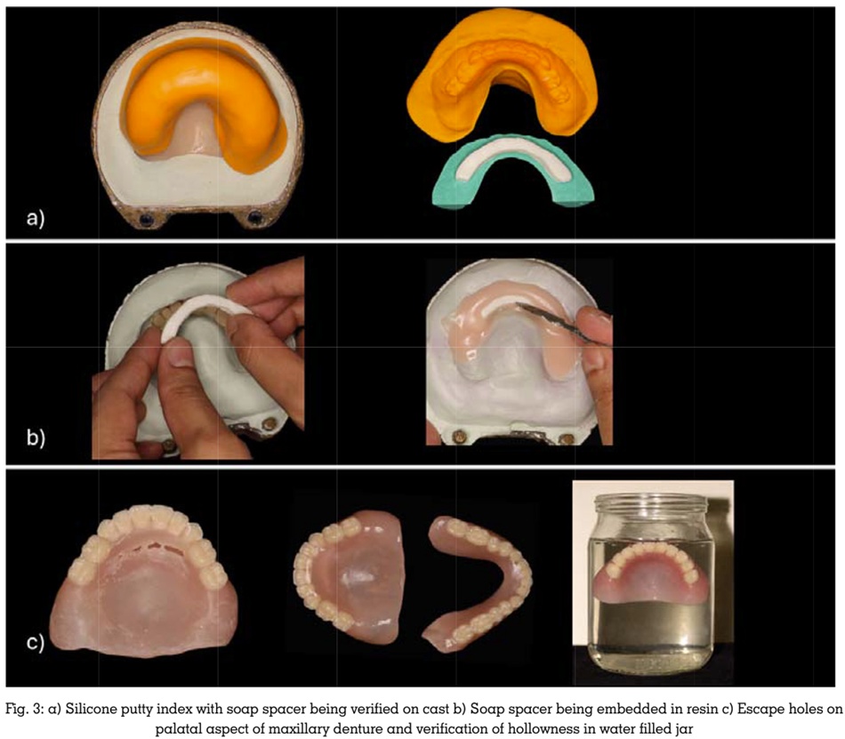
A fluid, early-dough mix of heat-polymerizing
polymethyl methacrylate (PMMA) resin (Acryton-
‘H’) was adapted into the mold cavity. The custom soap spacer was precisely embedded within
the center of this resin pack [Figure 3b]. The
remaining dough-stage PMMA was immediately
packed over the spacer before the initial layer
transitioned into a late dough stage. This
ensured a seamless chemical bond between the
upper and lower halves of the resin, mitigating
future split-line failures.
The flask was compressed in a hydraulic press
and processed using a standard long heat-curing cycle. Following deflasking, escape holes were
drilled into the distal palatal aspect of the cured
denture. Warm water was injected through these
access holes to dissolve and thoroughly flush out
the internal soap core, creating a clean internal
void. The escape holes were subsequently sealed
using auto-polymerizing resin. The hollowness
and reduction in mass of the maxillary denture
were verified by placing it in a water vessel
and observing its buoyancy [Figure 3c]. The
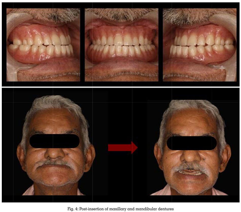
completed dentures were finished, polished, and delivered. Comprehensive post-insertion
hygiene and wear instructions were provided to
the patient [Figure 4].
Clinical evaluations were performed at 48
hours, 1 month, and 6 months post-insertion.
Minor pressure areas were identified on the mucosal surface during the initial 48-hour
follow-up and were relieved using a selective
grinding technique. At the 6-month recall, the
patient reported good retention and stability
in both arches, improved speech clarity, and a
significant improvement in masticatory efficiency
and comfort. The surrounding mucosa remained
healthy, with no signs of soft-tissue irritation or
ulceration.
Prosthetic
management of a completely
edentulous post-maxillectomy patient who has
undergone radiation therapy presents distinct
biomechanical hurdles. The loss of the premaxilla
shifts the prosthesis’s center of gravity posteriorly,
deprives it of crucial anterior skeletal support,
and alters the fundamental physics of denture
function.2 Additionally, irradiated tissues exhibit
reduced cell turnover and decreased micro
vascularity, making them highly susceptible to
chronic ulceration or osteoradionecrosis under
heavy, uneven structural loads.6
Reducing the overall mass of the maxillary
prosthesis is an established method to enhance
vertical retention and stability; minimizing the
gravitational pull reduces the dislodging forces
operating on the maxillary peripheral seal.[3]
While several materials can be used as three
dimensional placeholders during laboratory
processingincluding salt, sugar, and industrial
wax, soap offers unique technical advantages.
It
functions as an easily carved spacer that
maintains its structural volume under flask
compression, yet dissolves completely in warm
water, creating a clean internal void without
degrading the polymer matrix of the surrounding
acrylic resin. This approach aligns directly with
classic weight-reduction principles established
in prosthodontic literature.1,7
The mandibular arch presentation added further
complexity due to the combination of an anterior
hypermobile (“flabby”) ridge and advanced
posterior resorption (Atwood’s Order VI). This
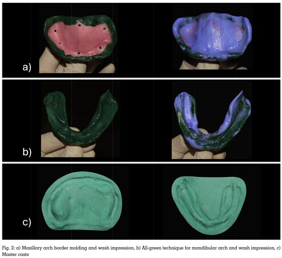
was successfully managed by applying the “all
green” border molding technique, which records
mucosal tissues under dynamic functional
loads, distributing masticatory forces across a
wider surface area to minimize localized stress
concentrations.8
While advanced 3D-printing and CAD/CAM
workflows are increasingly used to precisely
control internal wall thickness in modern max
illofacial prosthetics, conventional techniques
utilizing soluble spacers remain a clinically re
liable, accessible, and cost-effective standard
where advanced digital infrastructure is un
available.4,5,9
This case report describes a single clinical case with a relatively short follow-up period of six months. Objective quality-of-life parameters (such as validated OHIP questionnaires) and long-term mechanical fracture resistance of the hollowed PMMA base were not quantified. Further prospective clinical trials are required to validate the long-term performance of this protocol.
Rehabilitating
post-oncological, irradiated
edentulous patients requires creative modifica
tions to conventional prosthodontic protocols.
The soap-spacer technique used here provides a
simple, precise, and inexpensive method to man
ufacture a hollow maxillary complete denture.
By minimizing gravitational dislodgement and
reducing mechanical stress on compromised
supporting tissues, this approach represents a
highly promising alternative when surgical re
constructions or implant-retained options are
restricted by anatomical or financial constraints.
The patient provided all appropriate consent for
the publication of this case report. In the form,
the patient has given his consent for his images
and other clinical information to be reported
in the journal. The patientunderstands that his name and initial will not be published and due
efforts will be made to conceal his identity, but
anonymity cannot be guaranteed.