Implant rehabilitation in the anterior maxilla demands precise implant positioning and prosthetic planning to achieve optimal aesthetics and function. However, compromised implant angulation may adversely affect emergence profile, screw access positioning, and overall aesthetic outcomes. Customized CAD-CAM implant abutments provide a prosthetically driven solution for correcting angulation discrepancies and improving soft tissue support in such situations. This clinical report describes the aesthetic rehabilitation of a missing maxillary central incisor using an implant supported prosthesis with a CAD-CAM customized abutment. A 3.0 × 10 mm ADIN implant was placed in the edentulous site. During the prosthetic phase, compromised implant angulation necessitated the fabrication of a digitally designed customized abutment to achieve favourable emergence profile and aesthetics. Following CAD-CAM fabrication and prosthetic rehabilitation, satisfactory aesthetic integration and patient satisfaction were achieved. This report highlights the role of customized CAD CAM abutments in managing aesthetic challenges associated with compromised implant angulation in the anterior maxilla.
Key words: CAD-CAM, customized abutment, anterior implant, aesthetic rehabilitation, implant angulation, digital workflow
Replacement of a missing maxillary anterior
tooth with an implant-supported prosthesis is
considered a predictable treatment modality
for restoring aesthetics and function. However,
achieving an ideal aesthetic outcome in the
anterior maxilla remains clinically challenging
because of the high aesthetic demand and the
need for harmonious peri-implant soft tissue
architecture. Buccal bone resorption following
tooth loss or trauma may compromise implant
positioning and emergence profile, especially
in the aesthetic zone. In such situations, implant
placement is often performed using a two-stage
surgical protocol to facilitate osseointegration
and preserve peri-implant hard and soft tissue
stability.
Non-ideal implant angulation or labial implant
positioning can adversely affect the final
prosthetic outcome by producing unfavourable
screw access, compromised crown contour, and
inadequate soft tissue support. Conventional
stock abutments may provide limited correction
in these cases and may not adequately restore the desired emergence profile. Recent advances
in CAD-CAM technology have enabled
fabrication of customized implant abutments
with improved precision, enhanced emergence
profile control, and superior aesthetic integration.
Customized CAD-CAM abutments allow
individualized prosthetic correction according
to implant angulation and peri-implant soft
tissue requirements, thereby improving the final
aesthetic outcome1.
This clinical report describes the aesthetic
rehabilitation of a maxillary anterior implant
with compromised angulation using a CAD
CAM customized abutment.
A 24 year old male patient presented to the
Department of Prosthodontics and Crown and
Bridge with a missing maxillary central incisor following previous trauma. The patient expressed
concern regarding compromised aesthetics and
requested a fixed replacement option. Clinical
examination revealed an edentulous space
in relation to the maxillary central incisor with
associated labial hard tissue deficiency (Fig. 1).
Adjacent teeth were clinically healthy and oral
hygiene status was satisfactory.
Radiographic evaluation using orthopantomo
gram revealed adequate bone height for im
plant placement (Fig.2). Based on the clinical
and radiographic findings, implant-supported
rehabilitation was planned. Due to the pres
ence of labial bone deficiency and the need to
achieve predictable osseointegration and soft
tissue healing, a two-stage implant placement
protocol was selected.
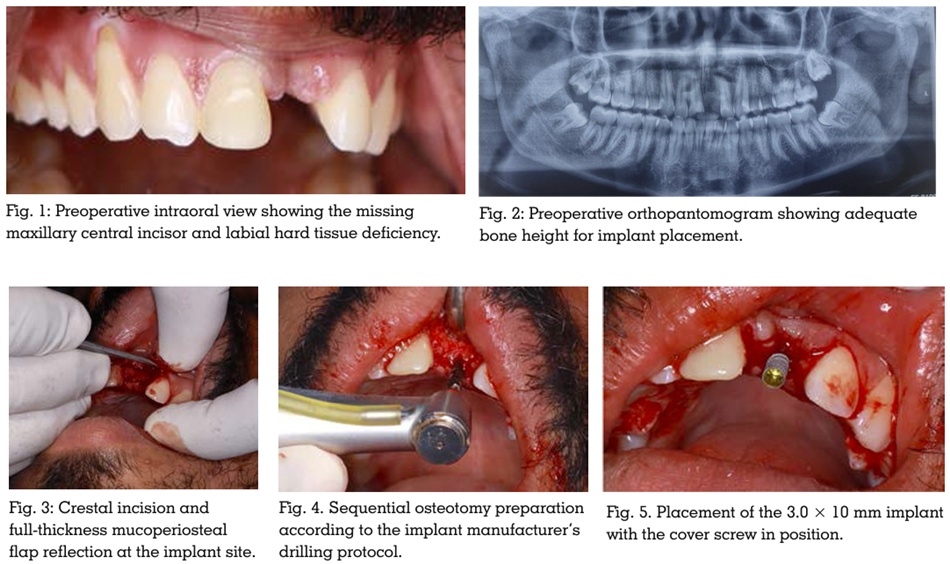
Under local anaesthesia and standard aseptic
precautions, a crestal incision was placed and a full-thickness mucoperiosteal flap was reflected
to expose the surgical site. Sequential osteotomy
preparation was performed according to the
manufacturer’s drilling protocol under copious saline irrigation. A 3.0 × 10 mm ADIN implant was
placed in the edentulous site with an insertion
torque of 45 Ncm, indicating satisfactory primary
stability. Following implant placement, a cover
screw was secured and the flap was repositioned and sutured to obtain primary closure (Fig. 3, 4,
5, 6). Postoperative instructions and medications
were provided to the patient. Healing during the
postoperative period was uneventful.
Following a healing period of four months,
second-stage surgery was performed and the
implant was exposed. Clinical evaluation during
the prosthetic phase revealed compromised
implant angulation with an unfavourable
emergence profile in the aesthetic zone. (Figure
7, 8). To overcome these limitations and improve
aesthetic integration, a customized CAD-CAM
abutment was planned. During the prosthetic
phase, an open-tray implant level impression
was made using elastomeric impression
material and transferred to the dental laboratory
for definitive prosthetic rehabilitation.
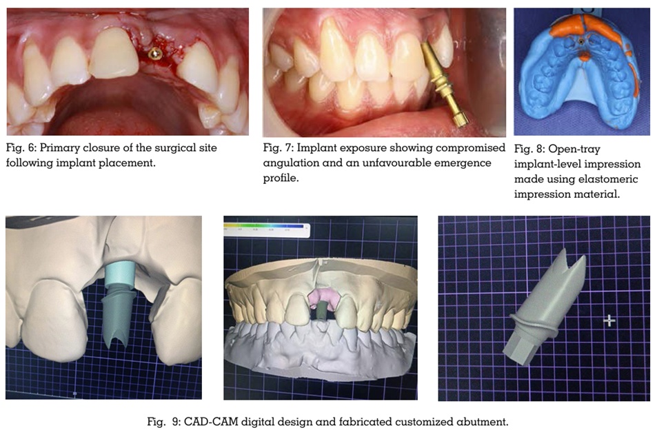
In the laboratory phase, the definitive cast was
digitally scanned after attaching a scan body
to transfer the implant position accurately into
the digital workflow. A customized implant
abutment was digitally designed using CAD
CAM software to improve the emergence profile
and compensate for compromised implant
angulation. The customized abutment was
subsequently milled and clinically evaluated for
fit, retention, and soft tissue support (Fig. 9).
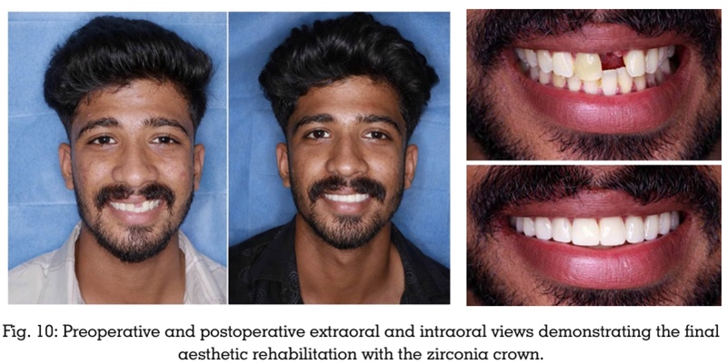
After satisfactory verification of the customized
abutment, a zirconia crown was fabricated and
cemented (Fig.10). The definitive prosthesis
demonstrated satisfactory aesthetics, functional
rehabilitation, and harmonious integration
with the adjacent dentition. The patient was
evaluated periodically after prosthesis delivery
at 3 weeks, 3 months, and 6 months follow-up
appointments. Clinical evaluation during follow
up revealed satisfactory peri-implant soft tissue
health, functional stability, and maintenance of
aesthetic outcome, with no evidence of prosthetic
or biological complications. The patient expressed satisfaction with the final treatment
outcome.
Achieving predictable aesthetic and functional
outcomes in anterior implant rehabilitation
requires precise coordination between surgical
implant placement and prosthetic reconstruction.
Implant position, angulation, and depth play a
decisive role in determining the final emergence
profile, peri-implant soft tissue contour, and long
term biomechanical stability. Mal-positioned
implants in the aesthetic zone present significant
restorative challenges because even minor
deviations in implant angulation can adversely
affect crown contour, gingival architecture,
phonetics, and occlusion. Previous studies have
emphasized that rehabilitation of malaligned
implants often requires customized restorative
approaches to achieve acceptable aesthetic
integration and prosthetic harmony2.
In the present case, the implant exhibited
compromised angulation and an unfavourable
emergence profile following second-stage
surgery. Conventional stock abutments were
unable to adequately compensate for the implant
trajectory because of their standardized form
and limited corrective capability. Bhavana et al.
reported that prefabricated abutments may not
provide sufficient correction in implants placed
outside the ideal restorative axis and highlighted
the advantages of customized abutments in such
situations3. Therefore, a customized CAD-CAM
abutment was selected to redirect the prosthetic
axis and establish improved aesthetic harmony
with the adjacent dentition.
Customized implant abutments have become
an important treatment option for rehabilitation
of mal-positioned implants, particularly in the
anterior maxilla. Customized abutments permit individualized contouring of the transmucosal
profile, improved soft tissue support, correction
of implant angulation, and better distribution
of occlusal forces. Cavallaro and Greenstein
discussed the practical application of angled
implant abutments in correcting implant
divergence and improving prosthetic alignment
in aesthetically demanding situations4. Similarly,
Sikri and Sikri emphasized that customized
and angled abutments can conservatively
manage compromised implant positioning while
improving aesthetics and function without the
need for implant removal5.
The introduction of CAD-CAM technology
has significantly improved the precision and
predictability of implant prosthodontics. Digital
workflows minimize laboratory inaccuracies
associated with conventional waxing and casting
procedures while improving marginal adaptation
and reproducibility. Barros et al. demonstrated
that CAD-CAM customized abutments allow
accurate correction of implant angulation
and facilitate fabrication of restorations with
improved biomechanical and aesthetic outcomes
in mal-positioned implants6. In the present case,
the digital workflow facilitated fabrication of a
customized abutment that successfully corrected
the prosthetic emergence profile and harmonized
with the surrounding dentition.
Peri-implant soft tissue management is another
important determinant of aesthetic success in
anterior implant rehabilitation. The contour
and emergence profile created by the abutment
significantly influence gingival adaptation,
papillary support, and mucosal stability. Jivraj
and Chee emphasized that proper treatment
planning in the aesthetic zone is essential for
achieving harmonious peri-implant soft tissue
architecture
and natural-looking implant
restorations7. Verma et al. reported that patient
specific customized abutments provide improved soft tissue support and natural gingival contours
in implants with compromised positioning8.
Similarly, the customized abutment in the
present case aided in achieving favourable peri
implant soft tissue adaptation and improved
crown contour despite the compromised implant
angulation.
Biomechanical considerations remain critical
in implants restored with angled or customized
abutments. Improper implant angulation can
increase stress concentration at the implant
abutment interface and may predispose the
restoration to complications such as screw
loosening, component fracture, and peri
implant bone loss. Pathak et al. noted that
excessive implant angulation and rotational
discrepancies may compromise screw joint
stability and long-term implant success. Long
term clinical success of angulated abutments
has nevertheless been reported by Sethi et al.,
who demonstrated favourable outcomes when
appropriate prosthetic planning and occlusal
control were maintained9. Therefore, careful
occlusal adjustment and elimination of excursive
interferences were performed in the present case
to minimize unfavourable loading conditions.
Comparative studies have demonstrated that
customized CAD-CAM abutments exhibit
superior marginal fit, improved emergence
profile, enhanced aesthetics, and better peri
implant tissue response compared to stock
abutments. Târtea et al. concluded that
customized CAD-CAM abutments provide
greater prosthetic accuracy and improved peri
implant tissue adaptation because of their
individualized design characteristics10. These
findings support the treatment strategy employed
in the present case.
Management of mal-positioned implants
using customized abutments and screw-retained restorations has also been reported
to provide predictable rehabilitation while
maintaining retrievability and facilitating
long-term maintenance11. In the present case,
the customized CAD-CAM abutment enabled
successful aesthetic and functional rehabilitation
without the need for additional invasive surgical
intervention.
Although customized CAD-CAM abutments
provide several clinical advantages, long-term
follow-up remains essential because restorations
supported by compromised implant angulation
may still be susceptible to biomechanical
complications over time. In the present
case, follow-up evaluations at three weeks,
three months, and six months demonstrated
satisfactory peri-implant soft tissue health,
stable prosthesis, absence of screw loosening,
and favourable patient satisfaction regarding
aesthetics and function.
Customized CAD-CAM abutments provide a
predictable and conservative treatment option
for rehabilitation of mal-positioned anterior
implants. In the present case, the customized
abutment successfully corrected the prosthetic
emergence profile, improved peri-implant soft
tissue adaptation, and achieved satisfactory
aesthetic and functional outcomes without
additional surgical intervention. Regular follow
up demonstrated stable peri-implant tissues and
favourable patient satisfaction.