
Aim: To compare the accuracy of intraoral scanners
and conventional impression techniques in recording
gingival sulcular depth and to evaluate their clinical
implications.
Materials and Methods: The prospective study
involved the collection of 20 samples comprising
10 anterior central incisors and 10 posterior molars.
Impressions for each tooth were recorded using
polyvinyl siloxane material with 3M Astringent
Retraction Paste and scanned using a desktop
scanner to acquire the STL files (Group I). The STL
files of the master cast models made from these
impressions were also acquired (Group M). STL
files were acquired using intraoral scanning after
retracting the gingiva (Group IOS). The deviation
in sulcular depth measurement was assessed by
superimposing all STL files using EXOCAD software.
Results: The mean deviation of the master cast group
(M) was found to be 0.0661 ± 0.0349 mm, whereas
the intraoral scanner group (IOS) had a greater
deviation at 0.1062 ± 0.1473 mm. The accuracy of the
conventional technique (93.39%) was significantly higher than the accuracy of the intraoral scanner
(89.38%). Nonetheless, there was no statistical
difference observed among both groups (p = 0.41).
Conclusion: In light of the results obtained in this
study, intraoral scanners are not as efficient as
the master cast in comparison to conventional
impressions in obtaining the sulcular depth. Despite
the greater accuracy shown by the conventional
approach, both methods are similar in their clinical
efficiency. Intraoral scanners can be viewed as an
effective clinical choice, but conventional impressions
remain the most reliable approach.
Key words: Intraoral scanner, conventional impression, sulcular depth, gingival retraction, accuracy
The longevity of restorations is influenced
by many factors. The final accuracy is often
influenced by the type of impression making,
setting accuracy, material flow, temperature,
humidity, mixing, disinfection, and pouring
time.1-3 When taking an impression, the gingival margin should be clear and accessible so that
the impression material can flow adequately.
Additionally, the gingival sulcus needs to be
sufficiently wide. The gingival displacement
technique’s inefficiency is the main cause of the
faulty marginal detail record2. Capturing the
gingival sulcular depth is one of the most crucial
steps in both traditional and digital impression
production to guarantee a precise marginal and
internal fit.
Accurate measurement is crucial for periodontal
assessment and prosthodontic design. Gingival
retraction cords or materials that temporarily
alter the marginal soft tissues are frequently
used in the traditional impression procedure to
enlarge the gingival sulcus without damaging
the delicate light material. However, it has a
primary drawback of causing bleeding which
will compromise the scanning accuracy. With the
advancement of three-dimensional technology
and the rising use of intraoral scanners in dental
office it is necessary to assess the accuracy of
scanners in capturing the gingival sulcular
depth employing cordless displacement devices
for fixed dental restoration ensuring proper fit.
The aim of this study was to compare the
accuracy of intraoral scanners and conventional
impression techniques in recording sulcular
depth and evaluate their clinical implications.
The null hypothesis is that there would exist no
significant difference in the reliability of intraoral scanner with the analog impression in capturing
the sulcus depth of prepared tooth using cordless
displacement system.
Sample Size Estimation
This prospective study was conducted at the
department of Prosthodontics and crown and
bridge at Rajas Dental College and Hospital
after
getting approval from Institutional
Research Board. Consent was asked from every
patient, explaining the study and its purpose.
Total study of 20 subjects was selected of which
10 posterior teeth (molars)and 10 anterior teeth
(Central Incisor) are selected with the presence
of adjacent teeth (mesial and distal) without
any prosthetic restoration, caries, periodontal
problem and malalignment. The sample size
estimation was done using G Power Software.
Study Design
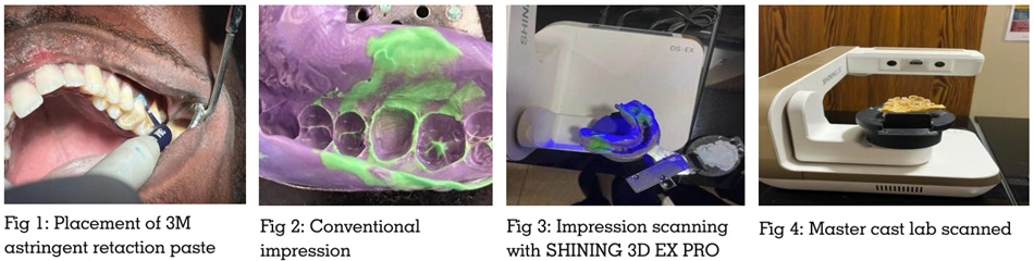
For every subject analog impression was taken
with polyvinyl siloxane impression material with
3M astringent retraction paste. The impression
was lab scanned (SHINING 3D EX PRO) and
STL file was created (Group I) (Fig: 1-3). The
master cast was poured from the impression and
the master cast was lab scanned (SHINING 3D
EX PRO) creating STL file (Group M) (Figure 4).
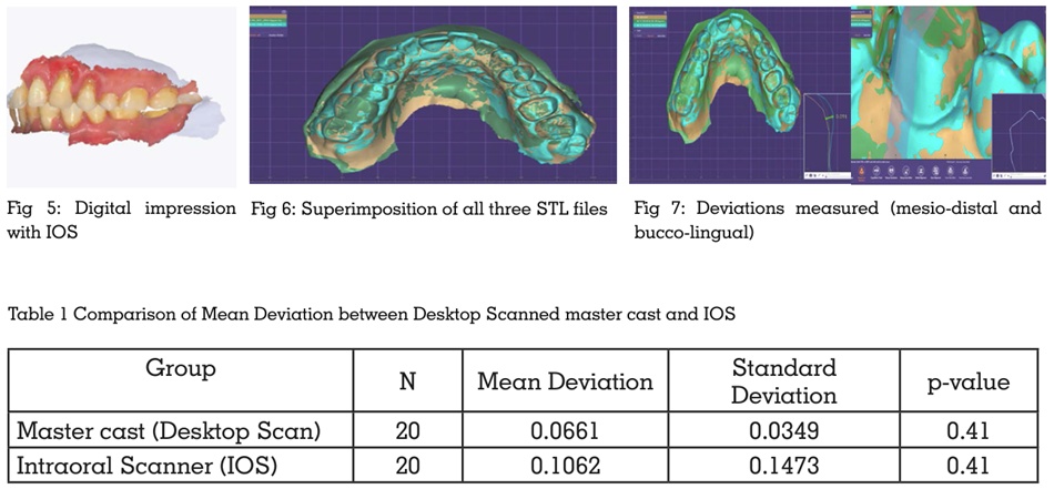
Digital impression was also taken with intraoral
scanner (CAMEO ELEGANT 3 IOS SCANNER) for which gingival retraction is done with 3M
astringent retraction paste and STL file is made
(GROUP IOS) (Figure 5)
Superimposition of all the three STL files are
done using EXOCAD software from which the
data will be acquired (Figure 6). By using the
Group I as control, deviation in recording the
gingival sulcular depth of Group M and Group
IOS measured (Figure 7)
A total of 20 dental samples, which included ten
posterior teeth molars and ten anterior central
incisors, were used to compare intraoral scanning
technique with conventional impressions for
recording sulcular depth. In this experiment,
conventionalimpression was taken using a
desktop scanner as the reference/control, and
then the deviation between master casts (STL
file M) and intraoral scans (STL file IOS) was
assessed based on the traditional impression
with the help of EXOCAD.
According to the findings of the analysis, the
mean deviation of the master cast (STL file
M) in comparison with impression was 0.0661
± 0.0349, whereas the mean deviation of the
intraoral scan (STL file IOS) compared with
impression was 0.1062 ± 0.1473. This proves the
fact that the master cast was more consistent
than the intraoral scanner.
As far as accuracy is concerned, the accuracy
of the impression approach was higher (93.39%)
than that of the intraoral scanning (89.38%).
Nevertheless, there were no statistically
significant differences between the two
approaches (p = 0.41). (Table 1)
Conclusively, even though the conventional
impression technique displayed a lower margin
of error and greater precision, both methods were
clinically equivalent, suggesting that intraoral
scanners may be deemed an equally clinically
viable option for recording sulcular depths.
Sulcular depth measurement is critical
for restorative and prosthodontic success,
influencing marginal fit, emergence profile, and
periodontal health. Conventional impressions
using polyvinyl siloxane (PVS) with retraction
paste are considered highly reliable for
capturing fine subgingival details, while
intraoral scanners (IOS) offer advantages such
as patient comfort, real-time visualization, and
a streamlined digital workflow. However, IOS
accuracy is limited by factors like moisture
control, soft tissue interference, and restricted
optical penetration.
In this study, the desktop-scanned impression
served as the reference. The master cast showed
better agreement with the control (0.0661 ±
0.0349 mm) compared to IOS (0.1062 ± 0.1473
mm), indicating greater variability with IOS.
Conventional impressions demonstrated higher
accuracy (93.39%) than IOS (89.38%), though
the difference was not statistically significant
(p=0.41),
suggesting comparable clinical
performance.
Previous systematic reviews indicate that while
IOS performs well in capturing hard tissues,
its accuracy decreases with mobile soft tissues
and deeper sulcular regions. Some studies
report superior or comparable accuracy of IOS
in specific applications, highlighting ongoing
advancements in digital technology.
Despite its limitations, IOS remains a promising
alternative; however, conventional impressions
continue to be more reliable, especially for deep
sulcular recording. The null hypothesis was not
rejected, as no significant difference was found
between the two methods, although conventional
techniques demonstrated greater precision.
Within the limitations of this study, the intraoral
scanner (IOS) was found to be less efficient
than the master cast when compared to the
conventional impression (control) in recording
gingival sulcular depth. The master cast
demonstrated closer agreement with the control,
indicating superior precision. However, with
ongoing advancements in IOS technology and
software, its accuracy is expected to improve
in the future. Despite these developments, the
conventional impression technique remains
the gold standard for recording deep gingival
sulcus.